
NCLEX-RN MI Wall Localization Cheat Sheet (Free Printable)
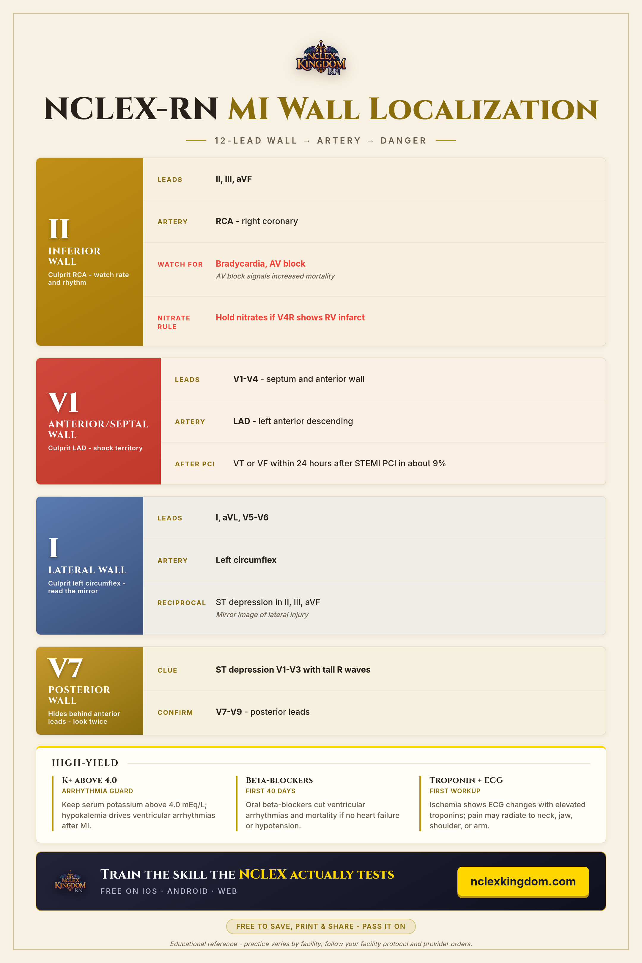
A 12-lead ECG does not just confirm an MI - it tells you which wall of the heart is involved and which artery is the likely culprit. That skill shows up on the NCLEX-RN because it drives bedside decisions: which rhythms to watch for, when to hold a medication, and what the first workup looks like.
This cheat sheet walks you through MI wall localization lead by lead - inferior (II, III, aVF), anterior/septal (V1-V4), lateral (I, aVL, V5-V6), and posterior (V7-V9) - and pairs each wall with its artery, from the RCA to the LAD to the left circumflex. It also flags the dangers tied to each territory, like bradycardia and AV block in inferior infarcts, plus pearls on potassium, beta-blockers, and the troponin and ECG workup. Read it once for the big picture, then quiz yourself on the lead-to-wall-to-artery chain. It is an educational reference, so always follow your facility protocol and provider orders.
Tap to open the full-size image in a new tab.
Educational reference - practice varies by facility, follow your facility protocol and provider orders.
Want to practice applying this under exam pressure? NCLEX Kingdom has thousands of NCLEX-style questions - start free in your browser.